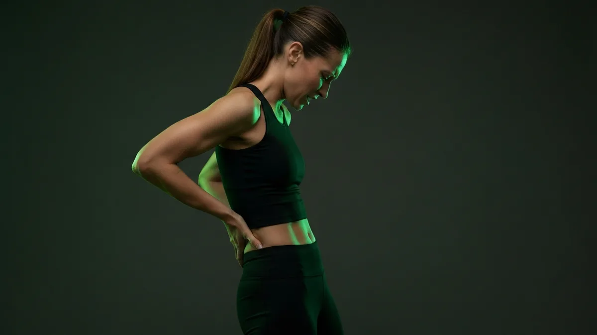
A rhomboid muscle strain occurs when the muscle fibers between the shoulder blade and spine are overstretched or torn — typically during a sudden pulling movement, heavy row, or forceful reaching motion. Symptoms include sharp localized pain along the medial scapular border, pain with shoulder blade retraction and protraction, and often protective muscle spasm. Most strains heal within 2-4 weeks with ice, gradual mobility restoration, and progressive strengthening.
How Rhomboid Strains Happen
The rhomboid major and minor are relatively small muscles that connect the medial scapular border to the thoracic spine. Despite their size, they bear significant load during any pulling movement — every barbell row, cable row, and pull-up loads these muscles during the scapular retraction phase.
Strains most commonly occur in three scenarios:
Sudden heavy pull. A jerky first rep on rows, catching a falling object, or an uncontrolled eccentric during pull-ups. The rhomboid is eccentrically loaded faster than it can produce force, and fibers tear. This is the classic "felt something pop" presentation.
Accumulated fatigue failure. High-volume rowing sessions where the rhomboids fatigue before the larger lats and traps. The final reps of the final sets produce the strain because the muscle has exhausted its capacity. This is the "didn't feel it until the next morning" presentation.
Pre-stretched overload. Reaching forward or across the body under load. The rhomboid is already in a lengthened position, and adding force in that stretched state exceeds the fiber tolerance. This happens during movements like reaching across the body while carrying weight, or forceful rotational movements.
Identifying the Strain
Location: Pain runs along the medial border of the scapula, between the shoulder blade and spine at approximately T2-T5 vertebral levels. The pain is usually on one side.
Aggravating movements: Squeezing the shoulder blades together (concentric rhomboid contraction), reaching forward with resistance (eccentric loading), deep breaths (the scapula moves slightly with rib cage expansion), and rotating the torso toward the affected side.
Relieving factors: Rest, gentle heat after the first 48 hours, and positions that place the shoulder blade in a neutral (non-retracted, non-protracted) position.
Distinguishing from other conditions: Rhomboid strains produce pain that is clearly muscular — worsening with specific muscle contractions and relieved by rest. If pain radiates around the rib cage, worsens with breathing deeply but not with shoulder movement, or is accompanied by fever, the cause may be something other than muscular strain and warrants medical evaluation.
Acute Treatment (Days 1-5)
Ice: 15-20 minutes every 2-3 hours for the first 48 hours. Ice the area directly over the medial scapular border. After 48 hours, transition to heat to promote blood flow and relaxation of the protective muscle spasm.
Modified activity: Avoid pulling movements, heavy lifting, and reaching overhead. Continue gentle daily activities. Walking is fine and beneficial — total rest delays healing.
Sleep positioning: Follow the guidelines in our rhomboid pain sleep guide — back sleeping with neutral thoracic support, or side sleeping with a pillow to prevent shoulder collapse.
Gentle mobility: Cat-cow stretches (on hands and knees, arching and rounding the thoracic spine) maintain thoracic mobility without directly loading the rhomboids. Shoulder rolls — slow, controlled circles — promote blood flow through the injured tissue.
Recovery Timeline
Days 1-5: Pain management. Ice, heat after 48 hours, gentle mobility. Avoid pulling and reaching under load.
Days 5-10: Begin light scapular exercises. Wall slides (arms against the wall, sliding up and down), light band pull-aparts at 50% of normal tension, and mid-back stretches. Pain should be noticeably decreasing.
Days 10-21: Progressive strengthening. Light reverse flies, face pulls with resistance bands, and cable rows at light weight with controlled tempo. Load increases week over week, never exceeding mild discomfort.
Days 21-35: Return to normal training. Reintroduce barbell rows and pull-ups at 50-60% of pre-injury loads. Progress by 10-15% per week. Full recovery is when you can perform all pulling movements at pre-injury loads without pain or apprehension.
Managing Trigger Points and Knots
Muscle knots frequently develop in the rhomboids following strain injuries. The injured tissue heals with adhesions and taut bands that produce ongoing tenderness even after the primary strain has resolved.
Self-release technique: Place a tennis ball or lacrosse ball between the affected area and a wall. Position the ball on the medial scapular border. Lean into the ball with moderate pressure and hold on tender points for 30-60 seconds until the tenderness begins to diminish. Move the ball systematically along the entire medial border from top to bottom.
Perform trigger point release daily during recovery and 2-3 times per week afterward for maintenance. Persistent trigger points that do not respond to self-release may benefit from professional manual therapy.
Preventing Re-Injury
Rhomboid strains recur when the underlying weakness or imbalance is not corrected. The three most effective prevention strategies:
Daily scapular retraction work. Band pull-aparts (3x15-20) performed every day build the endurance capacity that prevents fatigue-based strains. This is the single highest-yield preventive exercise for the rhomboids.
Controlled rowing eccentrics. On cable rows, emphasize a 2-3 second eccentric (letting the weight pull the arms forward in a controlled manner). Eccentric strength protects against the type of sudden stretch that causes acute strains.
Pull-to-push volume ratio. Maintain at least a 1.5:1 ratio of pulling to pressing volume in your training. This prevents the anterior dominance that overloads the rhomboids by constantly pulling the shoulders forward.
For the underlying anatomy, see our rhomboid muscle anatomy guide. For chronic rhomboid pain patterns, the postural correction and strengthening approach differs from acute strain management.